

ROTATOR CUFF TEARS
The rotator cuff is a group of four muscles around shoulder joint and provides stability to shoulder. Shoulder joint is a ball and socket type of joint in which the head of the humerus (upper arm bone) is the ball and glenoid of the shoulder blade is the socket of joint.
The rotator cuff is a group of four muscles:

- Supraspinatus is mainly responsible for shoulder abduction means lifting of the arm.
- Infraspinatus is responsible for external rotation of the arm at 0 degrees of abduction.
- Teres minor is responsible for external rotation of the arm at 90 degrees of abduction.
- Subscapularis balances the arm abduction and internal rotation of the shoulder.
Rotator cuff tears may involve single tendon or multiple tendons at a time. Most of the time rotator cuff tears are associated with AC joint pathology.
Mechanisms of injury
- chronic degenerative tears
- usually affects patients of older age groups
- it occurs as a result of chronic use and wear and tear
- most commonly involves the supraspinatus muscle followed by infraspinatus, teres minor muscles but it may extend anteriorly to involve the upper margin of subscapularis tendon in case of massive tears.
- acute avulsion injuries
- acute injuries are mainly subscapularis tears particularly seen in younger patients as a result of fall
- acute tears can also be seen in patients following a shoulder dislocation
- full thickness rotator cuff tears need to be repaired in young population and in throwing athletes.

Functions of rotator cuff
The rotator cuff muscles provide dynamic stability to shoulder joint by balancing the force couples that overshoulder joint in both the transverse and coronal plane.
- coronal plane
- the inferior rotator cuff muscles, posteriorly infraspinatus and teres minor, anteriorly subscapularis balance the superior moment created by the deltoid muscle.
- transverse plane
- Anteriorly subscapularis functions to balance the posterior directed moment created by infraspinatus and teres minor.
- this helps to maintain a stable fulcrum for glenohumeral motion.
- Main aim in treating rotator cuff tears is to restore this equilibrium in all planes of motion.
Cuff Tear Size
Small tears are – 0-1 cm
Medium sized tears are – 1-3 cm
Large tears are – 3-5 cm
Massive tears are more than 5 cm and involve multiple tendons.
Presentation and Symptoms
- gradual onset of pain which increases by overhead activities
- Night pain – when it is present, always indicates poor response to nonoperative management
- Loss or painful shoulder abduction
- Difficulty in combing hair, reach behind the neck, weakness in the arm.
Imaging of rotator cuff tears
- Radiographs
X rays are not commonly used. Anteroposterior view may show calcific tendonitis, calcification in the coracohumeral ligament. In case of chronic tears proximal migration of head of humerus is seen.
An outlet view may show a Type III acromion which is hook shaped, this can be a reason of rotator cuff tear.
- Arthrogram
This is also an uncommon procedure not used routinely. This is useful when MRI is contraindicated. If rotator cuff tear is present the dye will leak from glenohumeral joint into subacromial space.
- MRI
Best modality for diagnosing rotator cuff tears and pathology. It is also helpful in evaluating muscle quality, size, shape, and degree of retraction of tear. Degree of fat atrophy of muscle is best seen on sagittal images and also helps in ruling out biceps tendon pathology. Medial biceps tendon subluxation is indicative of a subscapularis tear.
- Ultrasound
This is another available option for imaging the rotator cuff tears. Main advantages are it allows for dynamic testing of rotator cuff, less expensive, easily available in most centers. But it is highly user dependent and needs lot of expertise to accurately diagnose the pathology.
Treatment
Important things to be considered before treatment
- Activity level and age of the patient
- Mechanism of tear (degenerative or traumatic avulsion)
- characteristics of tear like size, retraction of cuff and muscle atrophy
- Partial thickness tears vs. full thickness tears
- articular sided tears (PASTA lesion) vs. bursal sided tears
Nonoperative treatment
physical therapy, NSAIDS, and subacromial corticosteroid injections
Indications
- first line of treatment for most tears
- partial tears often can be managed with physiotherapy
- physical therapy with aggressive rotator cuff and scapular-stabilizer strengthening will help.
- subacromial injections are used if impingement thought to be major cause of symptoms.
- corticosteroids should not be used in full thickness rotator cuff tears.
Operative treatment
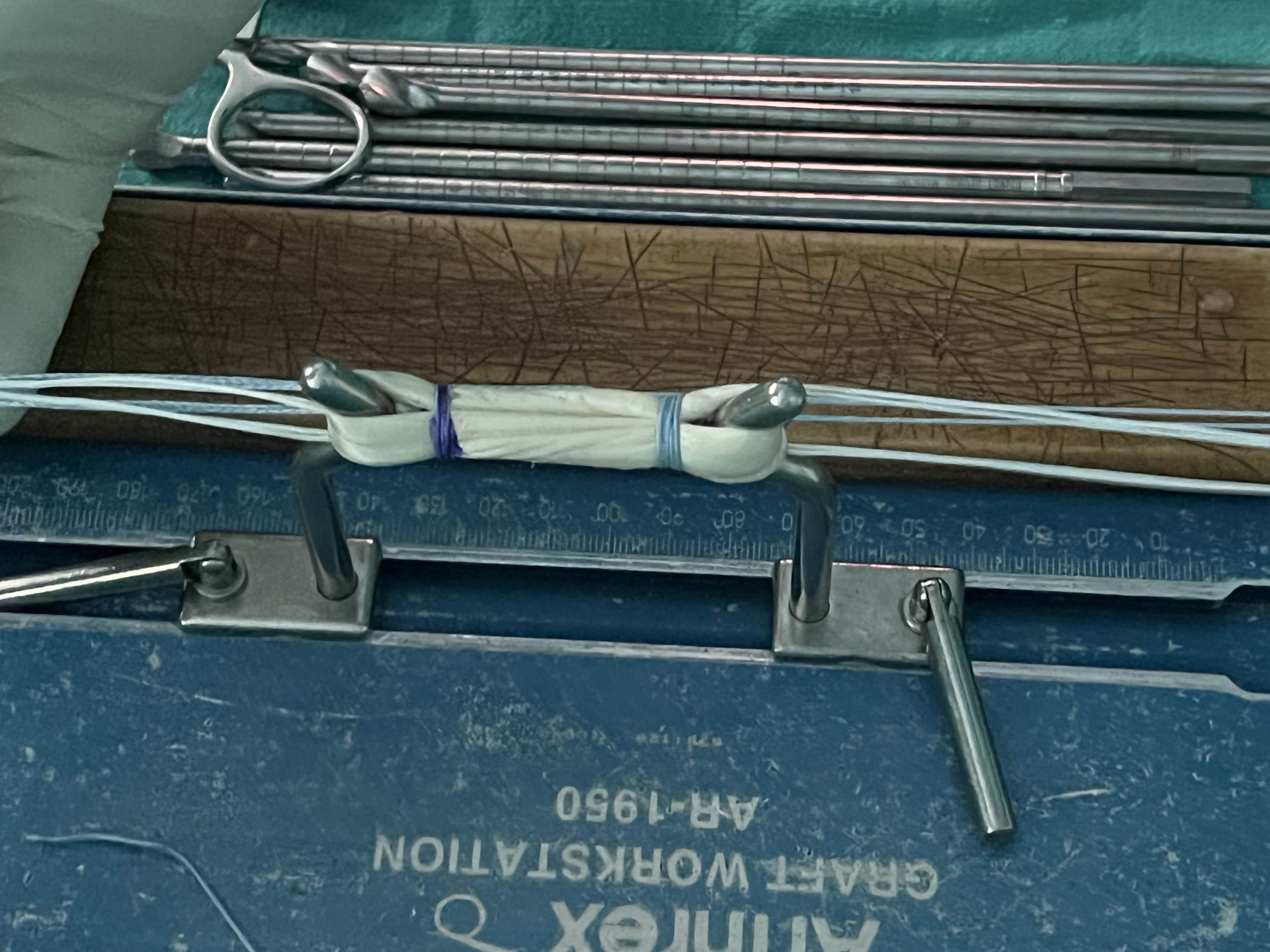
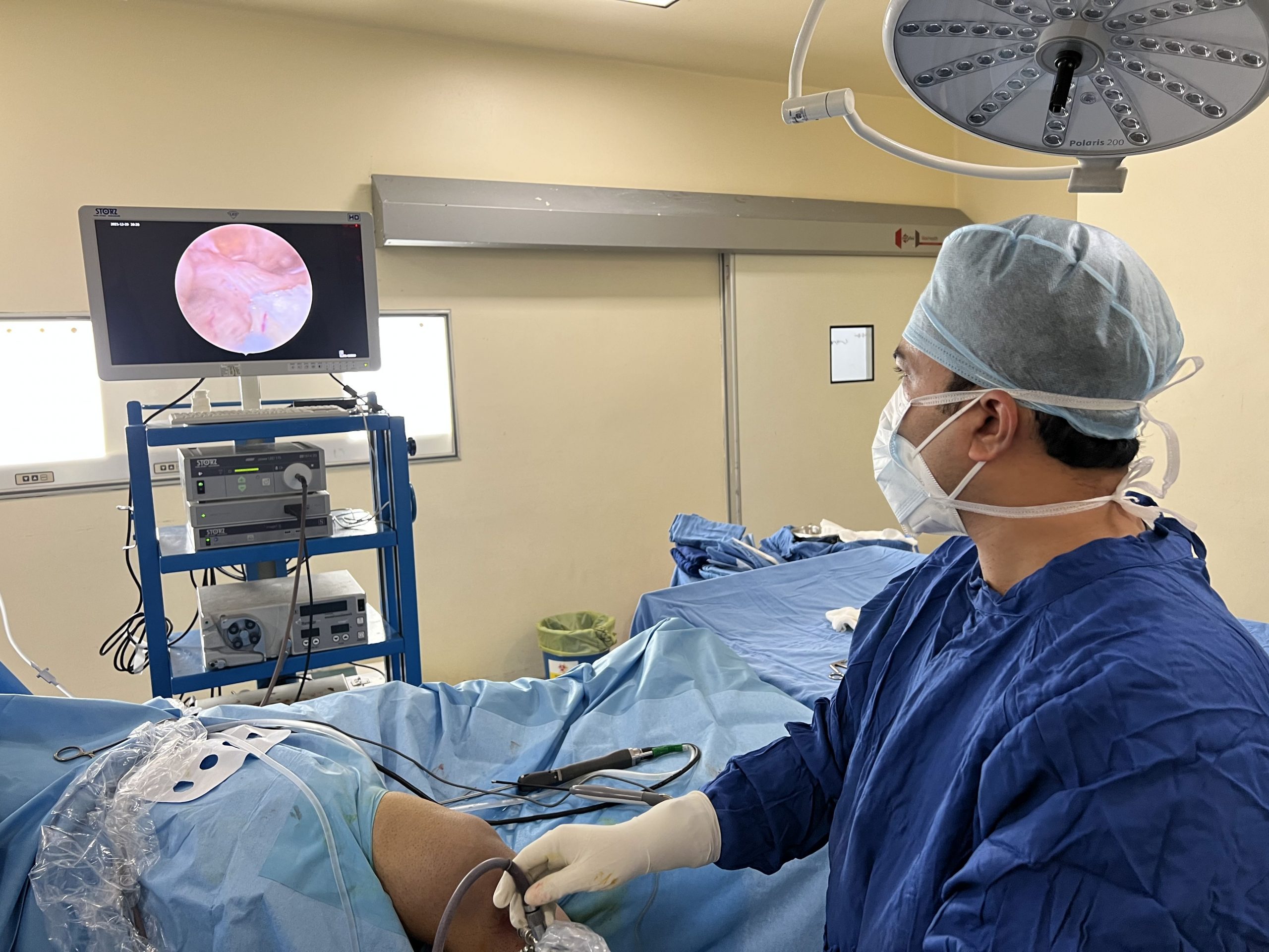
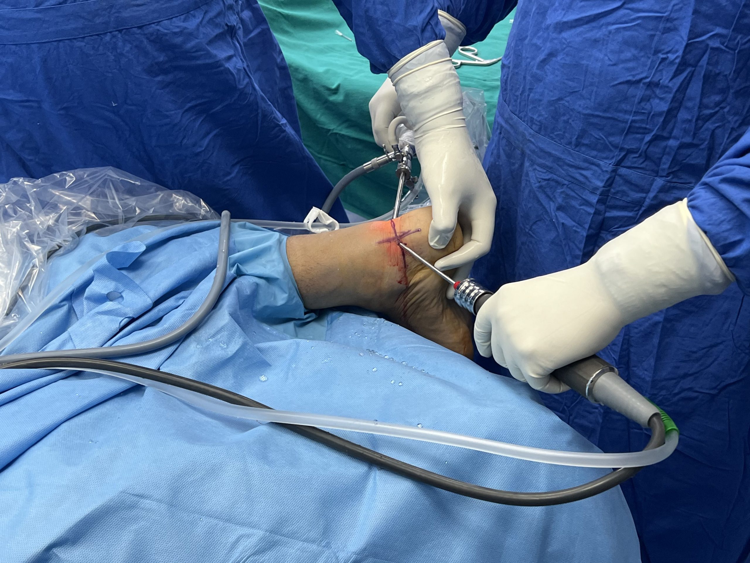
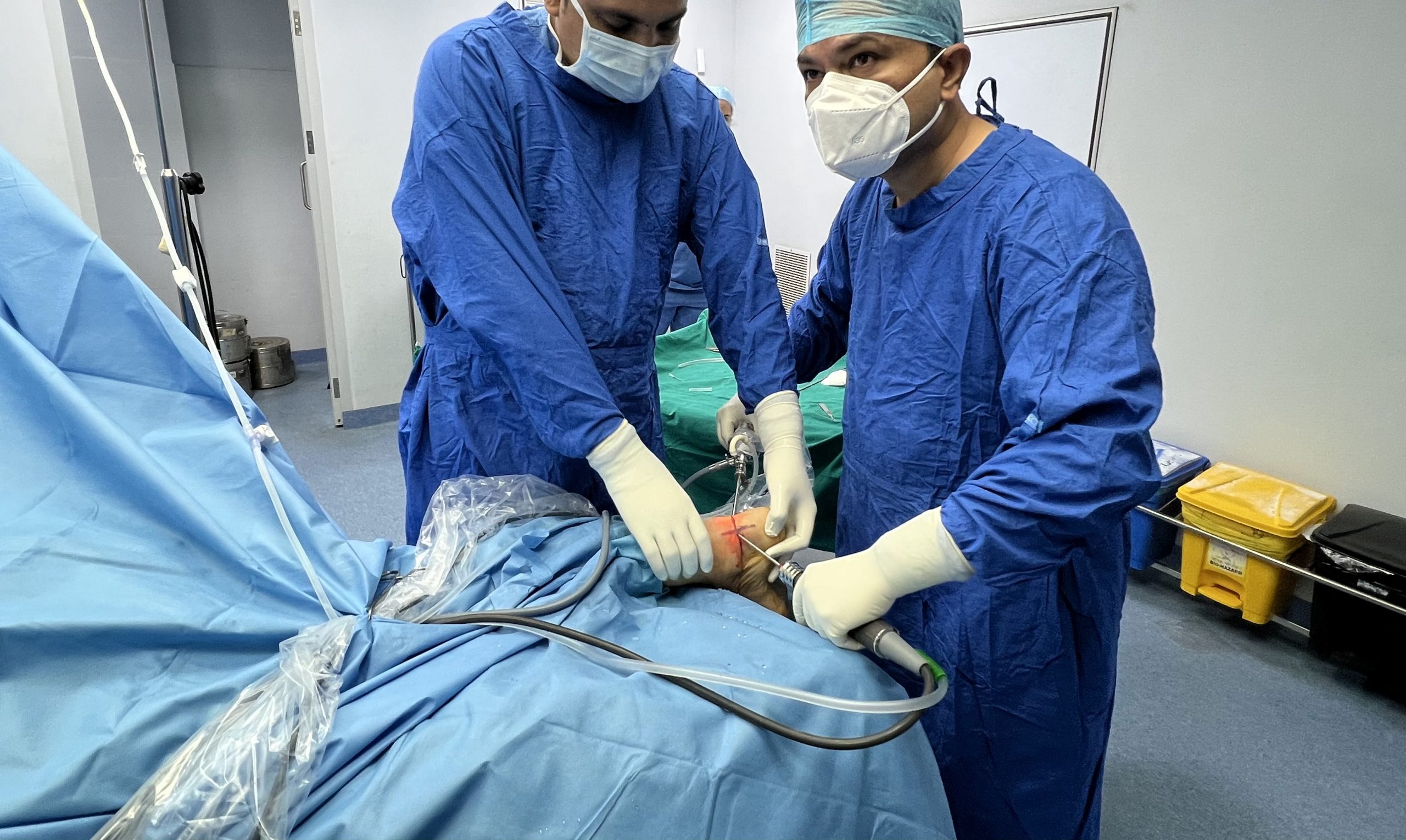
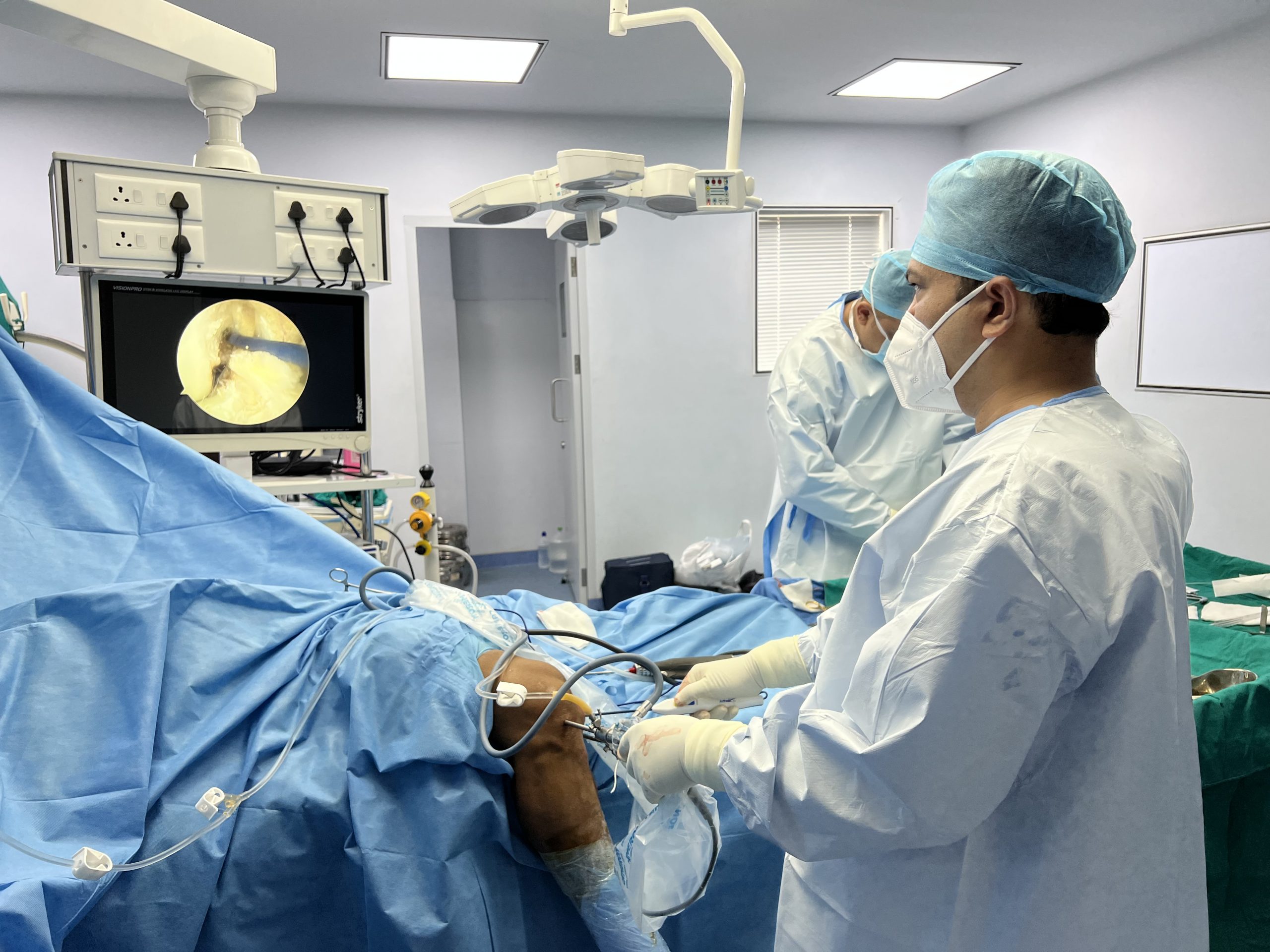
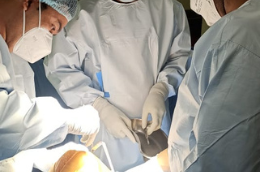
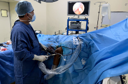
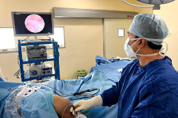
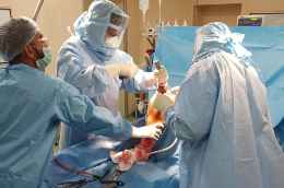
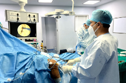
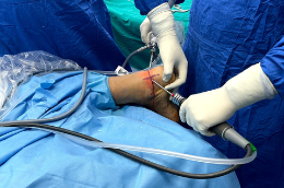
Arthroscopic rotator cuff repair
Indications
- Bursal-sided cuff tears more than 3 mm in depth
- Articular sided supraspinatus tears with more than 7 mm of bone exposed between the articular surface
- Rotator cuff tears those do not respond to conservative measures should be treated surgically.

Rate-limiting step for recovery is biologic healing of rotator cuff tendon to greater tuberosity, which is believed to take 8-12 weeks. Holes drilled in greater tuberosity are major source of vascularity to repaired rotator cuff.

Open or mini open rotator cuff repairs are not being used now a days because of latest advancements in arthroscopic procedures.


